ReaLiver Prototype
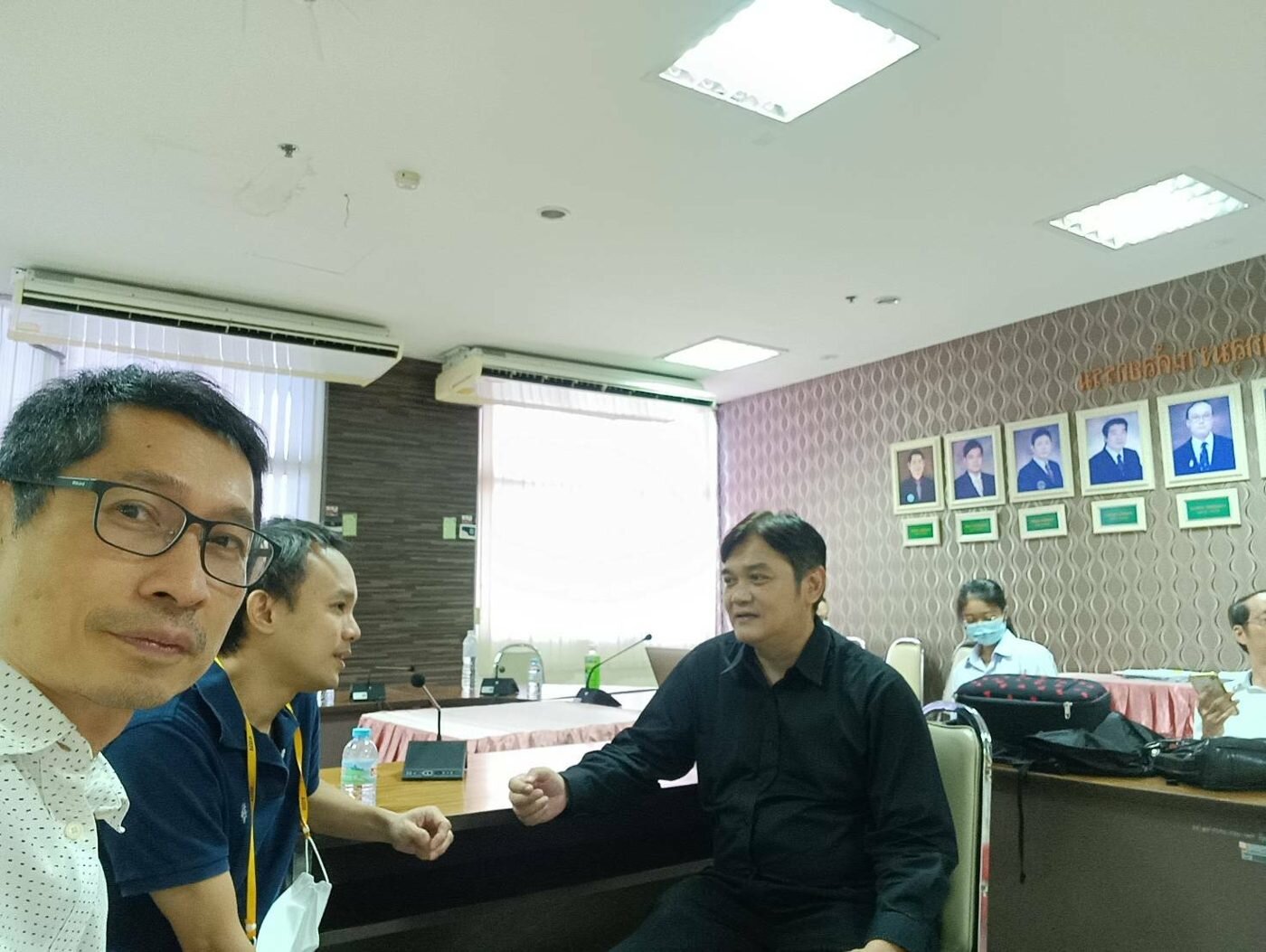
A prototype VR system for planning liver tumor resection, built from a routine CT scan and developed together with surgeons at a public hospital.
A prototype VR system for planning liver tumor resection, built from a routine CT scan and developed together with surgeons at a public hospital.
To remove a liver tumor, the surgeon must take out the tumor with a safe margin, keep enough healthy liver for the patient to recover, and avoid damaging the hepatic and portal veins. This is planned from stacks of 2D CT slices, which requires building a 3D picture of the tumor and vessels mentally — difficult to check, and difficult to share with the team.
Commercial 3D planning software exists, but its licence cost and IT requirements place it beyond most Thai public hospitals, where the majority of patients are treated. The CT data needed for 3D planning is already acquired for every case; what is missing is an affordable way to use it.
ReaLiver converts an existing CT scan into an interactive 3D model of the liver, its vessels, and the tumor, viewed in a VR headset. The surgeon sees depth and position directly, can rotate the liver and view it from any angle, and when a resection is proposed can see which vessels it would involve and how much liver would remain. The system is free for collaborating doctors and runs on consumer VR hardware with the hospital's own CT data.
Introduction video — the 3D liver resection planning workflow.
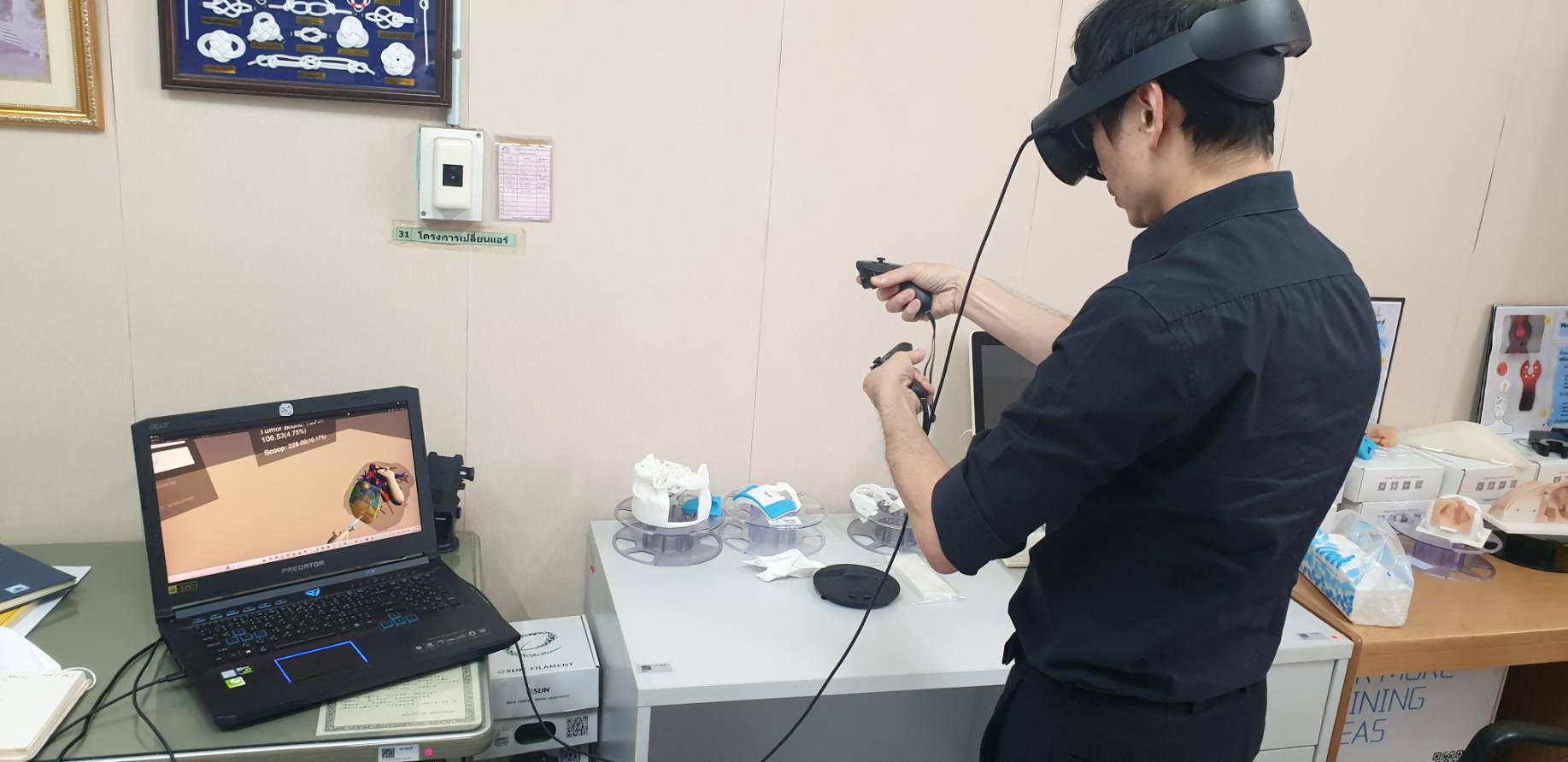
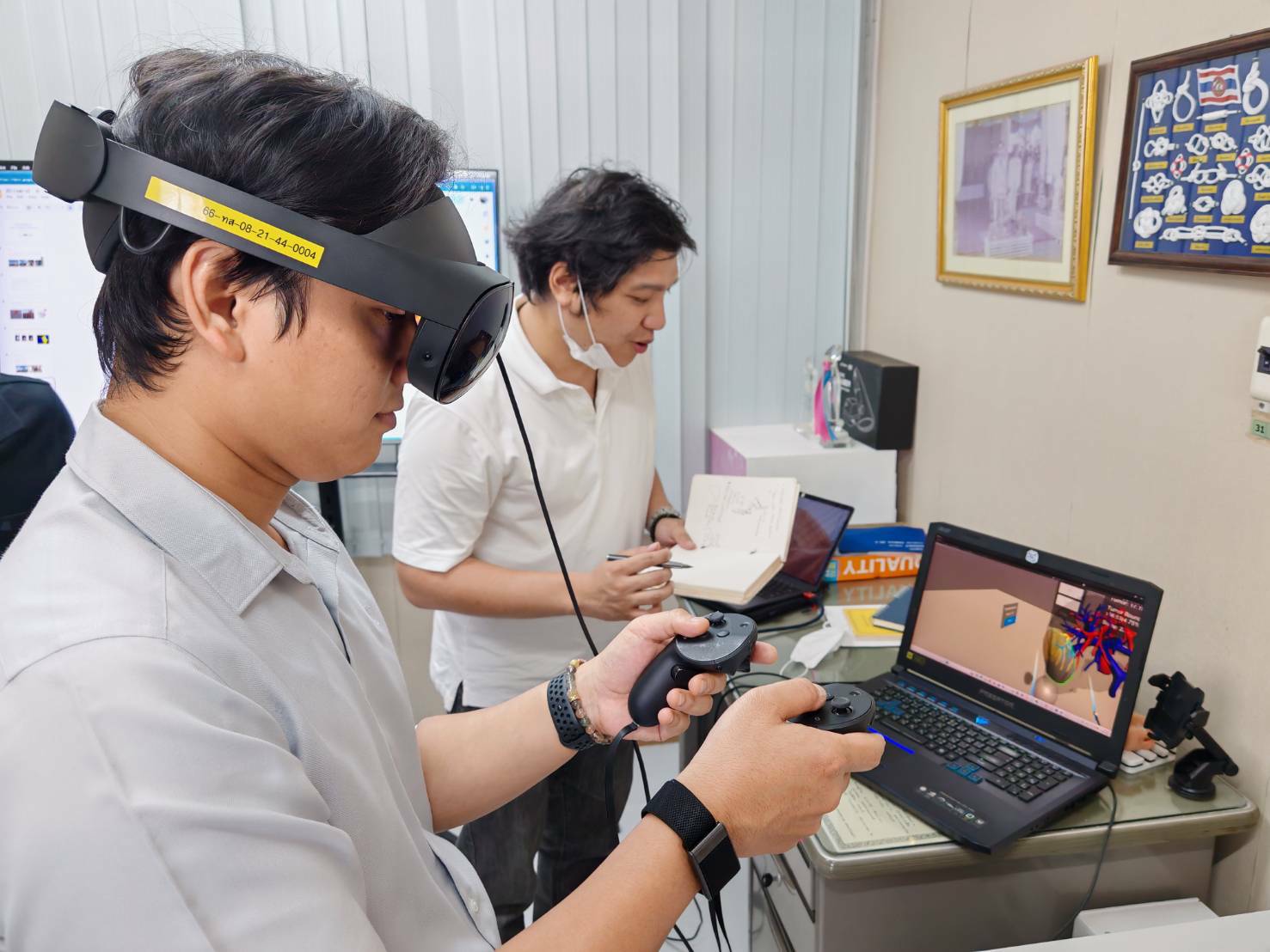
Demonstration of the VR application (version 3).
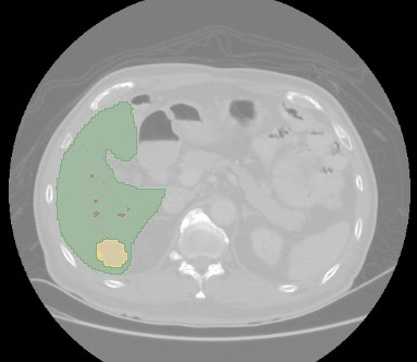
A routine CT scan is imported, and a deep-learning model separates the liver, the vessels inside it, and the tumor from the surrounding tissue. The liver is now segmented reliably. The vessels are harder, and the surgeons need the hepatic and portal veins shown as separate trees rather than as one tangle; this remains the main focus of the segmentation research.
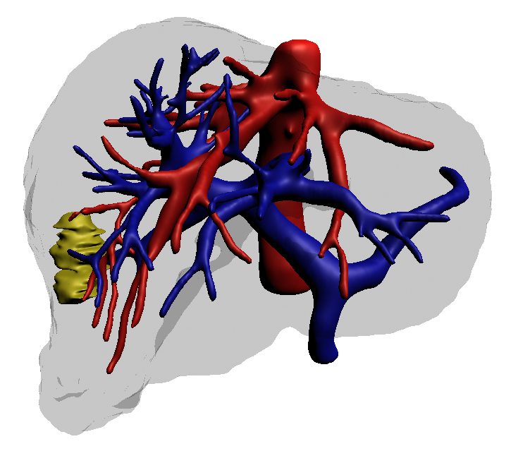
The segmented regions are converted into 3D surfaces. The hepatic and portal veins are built as separate structures so that the vessel network is legible inside a semi-transparent liver.
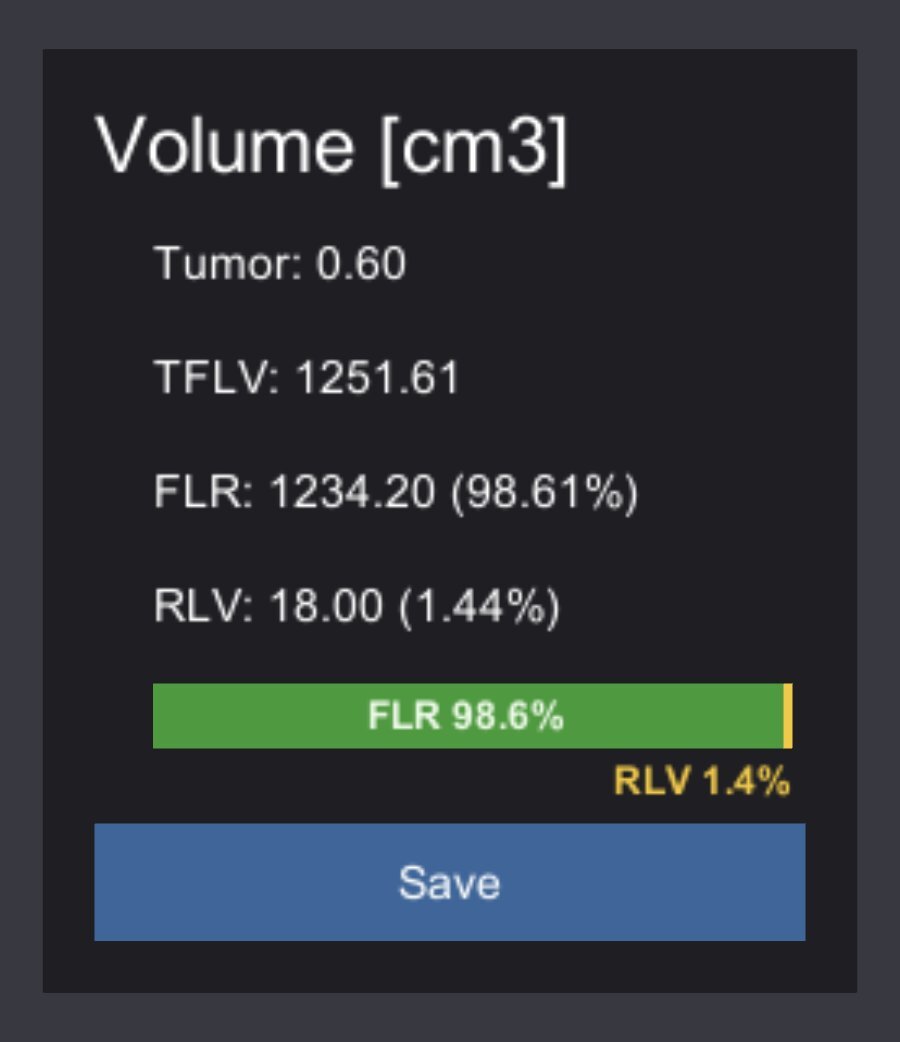
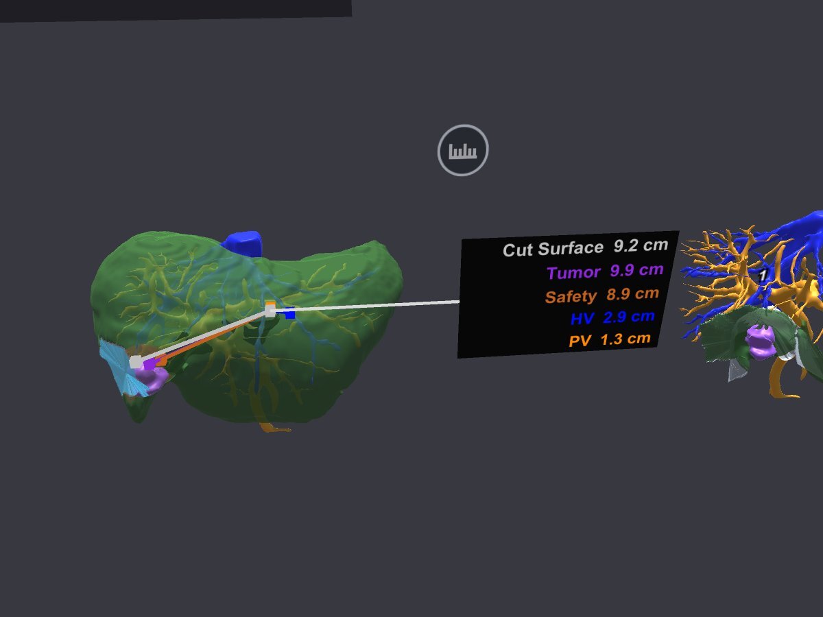
In the headset the surgeon rotates and inspects the model, then defines a resection region. The volume monitor updates the tumor volume, the resected volume, and the future liver remnant in real time. A 1 cm safety margin is drawn around the tumor, the remnant is checked against a 20–30% threshold, and any vessel the plan would cut is highlighted so the plan can be adjusted.
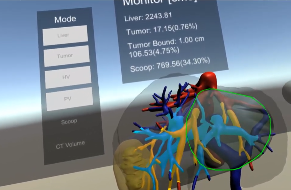
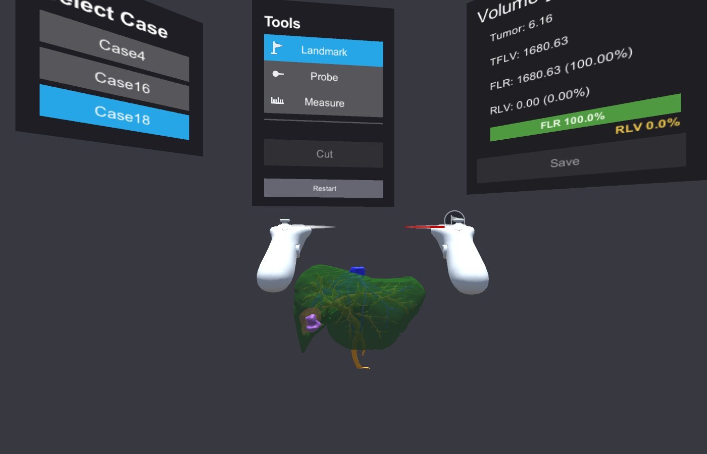
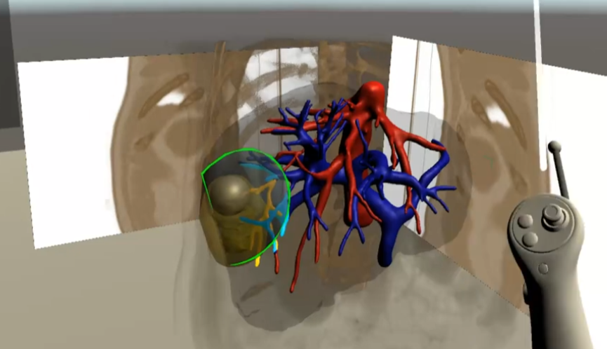
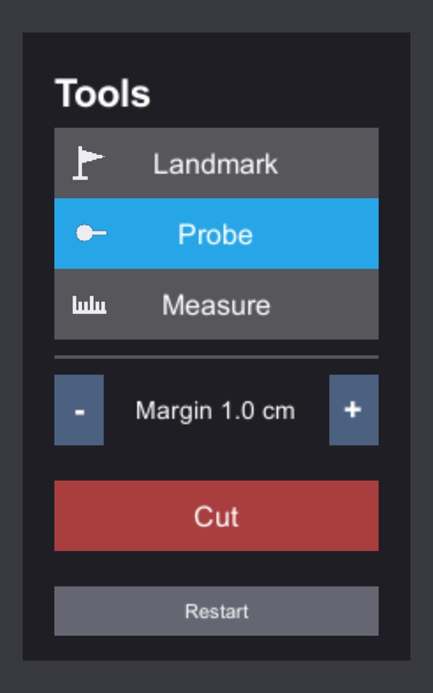
Probe. The surgeon places a control point on the liver surface, and the system projects a resection volume inward from that point to enclose the tumor and its margin. Moving the point shows which major vessels the approach would cross and how the future liver remnant changes, so the entry can be kept away from the hepatic and portal trunks while preserving healthy liver. The Probe is intended for non-anatomical resection of tumors near the liver surface, where the choice of entry point decides how much tissue is removed.
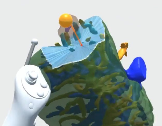
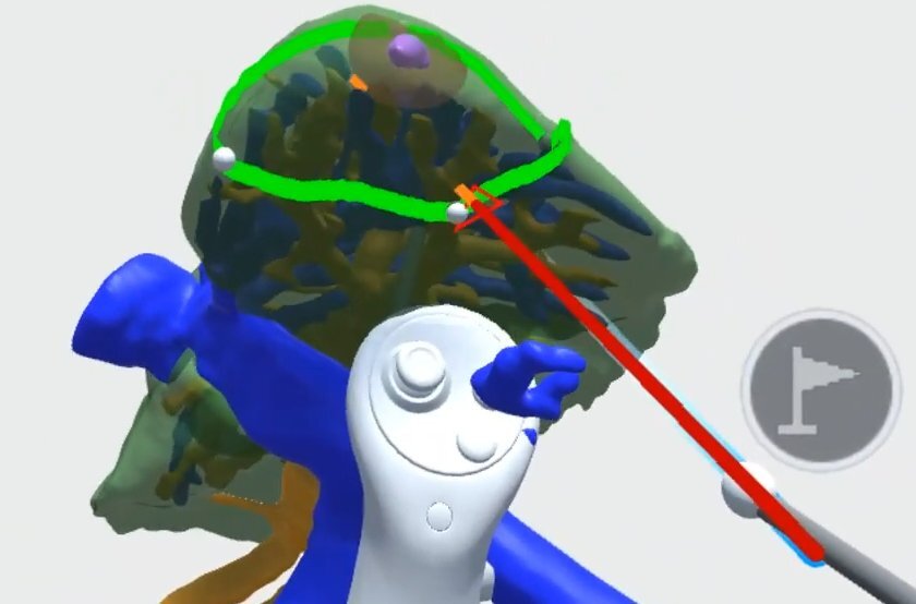
Landmark. The surgeon places a series of pins on the liver surface. A cut line is redrawn through the pins after each one is added, and a cut surface with adjustable depth divides the liver into the specimen and the remnant. This mirrors how a resection line is marked on the organ during surgery. The surgeons asked for this tool after using the Probe and consider it equally important.
Each plan is saved as a design with its margin, vessel involvement, and volumes recorded. Designs can be compared with each other and reviewed by the team before surgery.
Alongside the headset, a web-based interactive 3D report runs in any standard browser without installation. It pairs the rotatable model with linked axial, coronal, and sagittal CT views, resection-plan drawing tools, safety-margin display, and volume readouts, so that a plan can be prepared and notes recorded on any hospital computer.
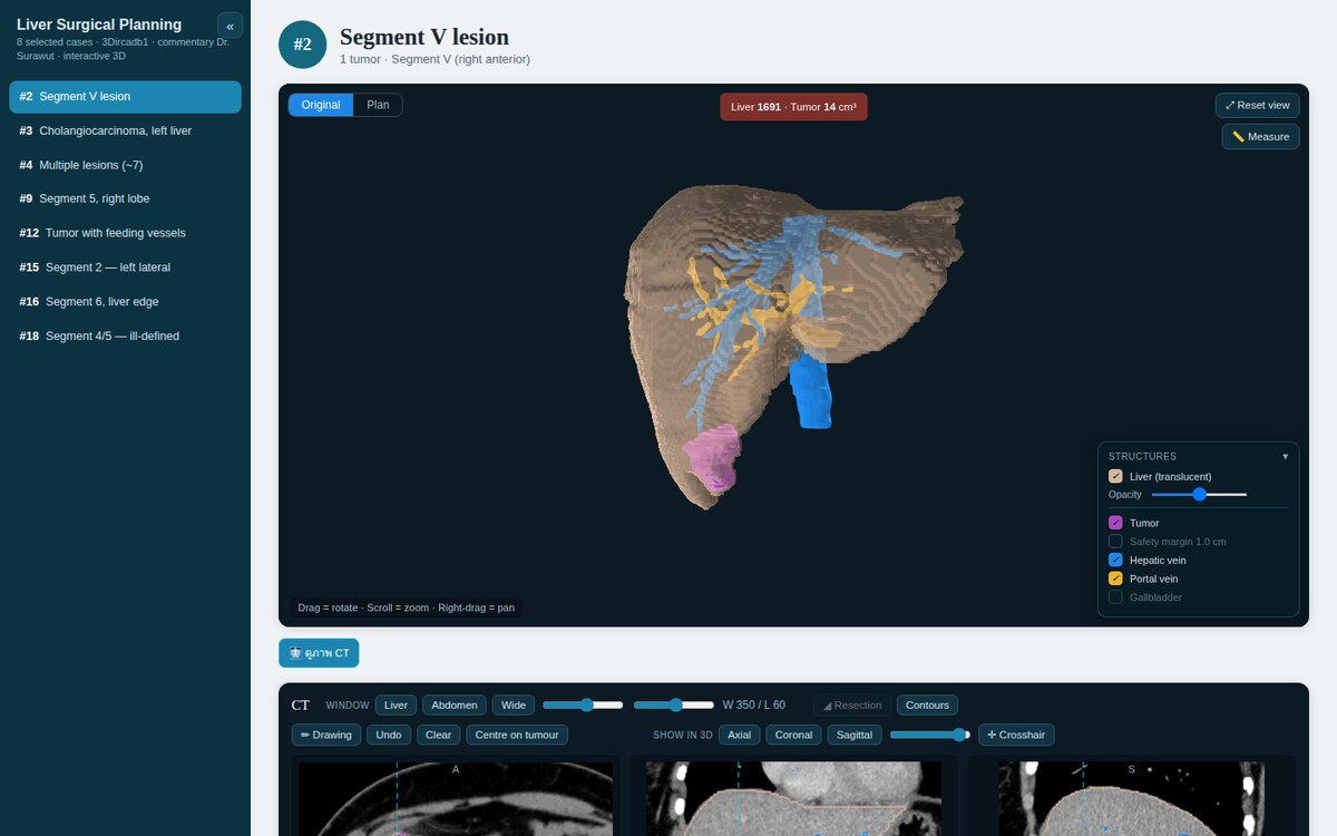
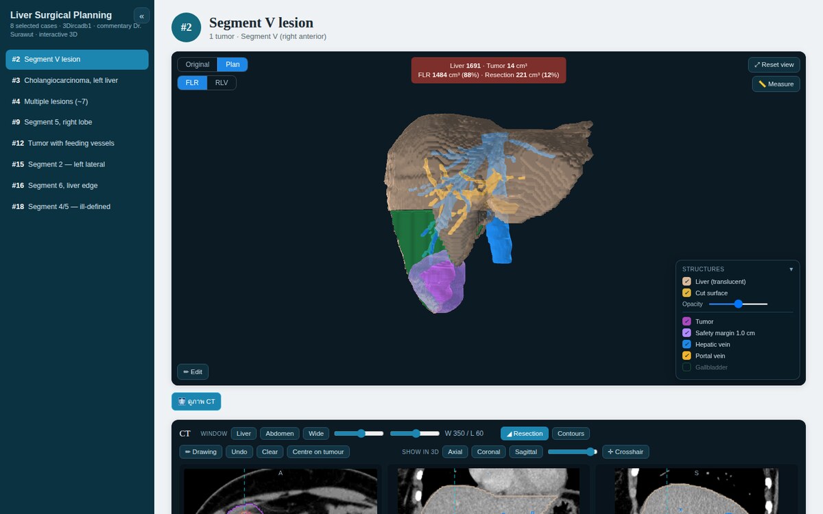
Note: the cases in this report are for testing and demonstration only. They use the public 3D-IRCADb dataset (IRCAD, France) — not patient cases from our partner hospital.
The work so far is prototyping. The cycle has been to gather requirements from the surgeons, build a prototype of what they asked for, let them try it, collect their feedback, and remove what they do not use. Both planning tools came out of this cycle. Over several feedback rounds the surgeons said the current prototype covers their requirements and is usable; this is informal feedback, and no formal evaluation has been carried out.
ReaLiver is a prototype. All test cases come from public datasets, no patient data has been used, and the system is not in clinical use.
Next steps are a formal evaluation with surgeons — for example planning time, workload, and plan quality — continued research on vessel segmentation, and the regulatory steps required before clinical use. A manuscript describing the planning technique has been prepared for journal submission. The same low-cost pipeline is intended to extend to other organs and other public hospitals.